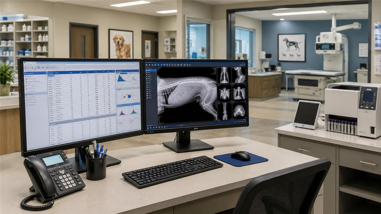
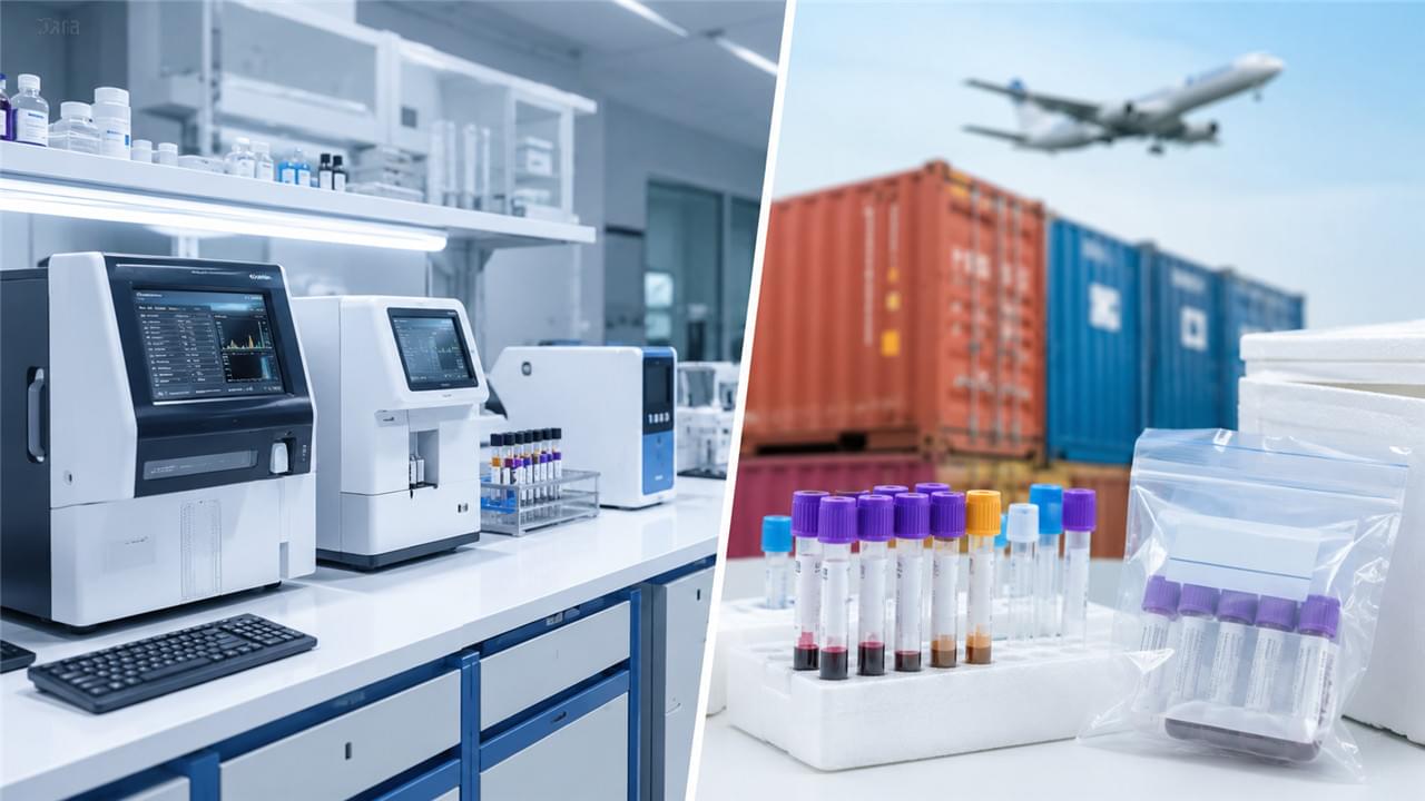
Veterinary diagnostic workflow — how samples flow from patient to result, how imaging gets reviewed, how data reaches the clinician at the right moment — determines whether your equipment investment improves outcomes or just decorates the building. Owning top-tier diagnostic equipment is necessary, but it’s nowhere near sufficient.
Diagnostic workflow optimization for veterinary hospitals is the operational discipline that closes the gap between equipment capability and clinical performance. This guide walks through the five problems that quietly drain diagnostic efficiency in most practices — and the practical fixes for each. For the full equipment overview, see our guide on Veterinary Diagnostic Equipment: From Basic Exams to Advanced Imaging.
🔗 Problem 1: Diagnostic Silos — Equipment That Doesn’t Communicate
Break the Silos That Slow Your Veterinary Diagnostic Workflow
In many practices, diagnostic equipment operates in isolation:
- Lab analyzers print results on paper or display them on a local screen
- Imaging systems store images on a local hard drive, disconnected from the clinical workstation
- Rapid test results get manually transcribed into patient records
Every one of these disconnections introduces delay, transcription error risk, and quiet information loss. The fix is veterinary LIS PACS integration at the practice level:
- LIS (Laboratory Information System): Connects analyzers directly to the practice management software (PMS), auto-populating patient records with lab results — manual transcription disappears entirely
- PACS (Picture Archiving and Communication System): Centralizes imaging storage (X-ray, ultrasound, dental) so any workstation in the clinic can pull up any study
- DICOM Worklist: Lets the PMS push patient data automatically to imaging equipment at study request, killing re-entry of patient information
Modern PMS platforms (ezyVet, VetSmart, AVImark) support LIS/PACS integration — prioritize integration capability when you select both your PMS and your diagnostic equipment. It’s the single highest-leverage improvement in veterinary diagnostic workflow, and the FAQ below explains why.
🧪 Problem 2: Sample Routing Delays
Speed Up Sample Flow in Your Veterinary Diagnostic Workflow
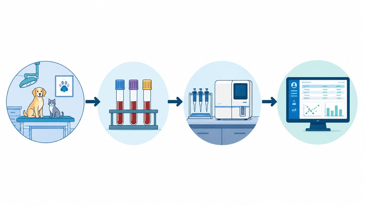
In high-volume practices, blood samples drawn in the exam room can sit there for 10–30 minutes before reaching the lab bench. At a practice running 40 consultations a day, that’s 6–20 hours of cumulative diagnostic delay every week — patients waiting, appointments stacking, decisions postponed.
Three fixes move the needle:
- Dedicated diagnostic technician: One nurse/technician assigned exclusively to sample processing during peak hours eliminates the gap between collection and analyzer run
- Satellite lab station near exam rooms: A micro-hematocrit centrifuge and automated analyzer in the treatment area (not a distant lab room) cuts transport distance and delay
- Standardized collection protocols: Training all staff on tube selection, fill volume, and mixing protocol reduces rejected samples and repeat draws — a silent win for veterinary diagnostic workflow efficiency
🖥️ Problem 3: Imaging Review Bottlenecks
Clear Imaging Queues in Your Veterinary Diagnostic Workflow
If only one clinician can review X-rays — because the imaging software lives on a single workstation — every radiograph study creates a queue, and queues turn into waiting-room pressure.
Open the flow with three changes:
- Multi-station PACS access: DICOM viewers on every clinical workstation (or cloud browser access) let multiple clinicians review studies concurrently
- Teleradiology access: For complex or uncertain findings, a teleradiology service (VetCT, Diagnostic Imaging Analysis Group) returns specialist interpretation within 2–24 hours — a massive confidence boost for referring practices
- Image reporting templates: Structured radiograph templates in the PMS (lung fields, liver size, skeletal assessment) create a consistent review checklist and better documentation
📋 Problem 4: Inconsistent Diagnostic Protocol Application
Standardize Protocols Across Your Veterinary Diagnostic Workflow
Without standardized protocols, each clinician applies a different diagnostic sequence to the same presentation. The result: inconsistent test ordering, under-utilized equipment (the ultrasound machine books out while routine abdominal screenings get skipped), and diagnostic gaps that only surface at clinical review.
Two solutions that actually stick:
- Disease-presentation pathways: Build clinic-level protocols for your top 10–15 presentations (“acute vomiting: CBC, chemistry, abdominal ultrasound”; “senior wellness: CBC, chemistry, UA, T4, BP”)
- PMS-triggered protocol prompts: Many modern PMS systems fire diagnosis-based reminders at case entry, prompting clinicians to order the indicated diagnostics — your veterinary diagnostic protocols become part of the software, not a laminated poster
🔧 Problem 5: Diagnostic Equipment Downtime
Keep Your Veterinary Diagnostic Workflow Running
An analyzer or imaging system out for repair doesn’t just cost revenue — it sends referrals out the door and breaks the rhythm of your veterinary diagnostic workflow. Both outcomes hurt.
Mitigate before failure:
- Preventive maintenance contracts: Schedule annual or biannual maintenance on all major systems; don’t wait for breakdowns
- Service level agreements: Require max 48-hour on-site response (24 hours for imaging systems) from providers
- Backup consumables: Never run below one week of reagent stock for in-house analyzers
- Loaner provisions: Negotiate loaner equipment for high-dependency systems (DR, ultrasound) in your service contracts
❓ Frequently Asked Questions
Q: What’s the most impactful single change a clinic can make to improve diagnostic workflow?
A: LIS integration between in-house analyzers and the practice management software. It eliminates manual result transcription, reduces errors, and puts results on any workstation instantly. Most modern analyzers support this at low or no additional cost beyond PMS configuration — the single biggest lever in veterinary diagnostic workflow.
Q: How do I get staff to consistently follow diagnostic protocols?
A: Compliance improves when protocols are embedded in the software workflow rather than enforced through reminders and meetings. PMS-triggered prompts (“Suspected UTI: recommend UA + culture”) at diagnosis entry remove the decision burden from individual clinicians and create a natural compliance path within your veterinary diagnostic workflow. Train-the-trainer sessions with one protocol champion per team also boost adoption.
Q: Is cloud-based PACS worth the ongoing subscription cost compared to a local server?
A: For most practices with 1–3 locations, yes. Cloud PACS eliminates local server hardware and maintenance, offers universal device access, built-in disaster recovery, and remote specialist access for teleradiology. The annual subscription (1,200–1,200–3,600 for most veterinary platforms) is typically justified by the overhead it removes — and it keeps veterinary diagnostic workflow running from anywhere.
✅ The Bottom Line
Diagnostic excellence is 30% equipment and 70% workflow. Start with veterinary LIS PACS integration, then fix sample routing, imaging review, protocols, and downtime — roughly in that order of impact. The WSAVA quality of care guidelines and AVMA practice management resources are excellent references for benchmarking your progress. And if you’re still deciding between in-house and external lab testing, our In-House Lab vs External Lab: Equipment Decision Guide covers that trade-off in depth.