Every successful surgery depends on far more than the skill of the surgeon. 🩺 Behind every sterile instrument and every clean incision is a carefully managed sterilization system that protects patients from avoidable infections. Veterinary surgical sterilization is not a single procedure—it is a continuous workflow that begins immediately after surgery and ends only when sterile instruments are safely opened for the next patient.
Even minor failures during cleaning, packaging, sterilization, storage, or documentation can increase the risk of surgical site infections (SSIs). Fortunately, these risks are largely preventable when veterinary hospitals implement standardized protocols, validated equipment, and routine quality monitoring.
In this guide, you’ll learn how to build an effective sterilization workflow, choose the right equipment, validate every sterilization cycle, and strengthen infection control in veterinary operating rooms. If you’re planning an entire surgical facility, our guide to Veterinary Surgical Suite Equipment: Systems, Standards & Selection provides a broader overview of surgical suite planning:
https://www.veteqpt.com/veterinary-surgical-suite-equipment
The Veterinary Surgical Instrument Sterilization Process: A Complete Workflow
Successful sterilization starts long before instruments enter an autoclave. 🔬 Every stage of the veterinary surgical instrument sterilization process contributes to patient safety, instrument longevity, and regulatory compliance.
The complete reprocessing cycle can be divided into five essential stages.
✅ 1. Immediate Point-of-Use Care
Immediately after surgery, blood and tissue should never be allowed to dry on surgical instruments. Moisture helps prevent biofilm formation, making subsequent cleaning significantly more effective. Delays at this stage often create contamination that becomes difficult to remove later.
🧼 2. Thorough Cleaning
Cleaning removes visible debris before sterilization begins. Depending on the workload, clinics may use manual cleaning, enzymatic detergents, ultrasonic cleaners, or automated washer-disinfectors.
This step is critical because steam cannot reliably sterilize instruments that still contain organic residue.
🔍 3. Inspection and Functional Testing
Every instrument should be carefully examined before packaging. Staff should check for:
- Corrosion
- Loose joints
- Damaged cutting edges
- Misaligned jaws
- Surface cracks
- Proper instrument movement
Removing damaged instruments before sterilization prevents unexpected failures during surgery while extending the lifespan of surgical sets.
📦 4. Packaging and Identification
Packaging serves as the sterile barrier that protects instruments until use. Each package should include:
- Chemical indicator
- Sterilization date
- Load number
- Operator identification
- Expiration or event-related tracking information
Proper packaging also allows complete steam penetration while maintaining sterility during storage and transportation.
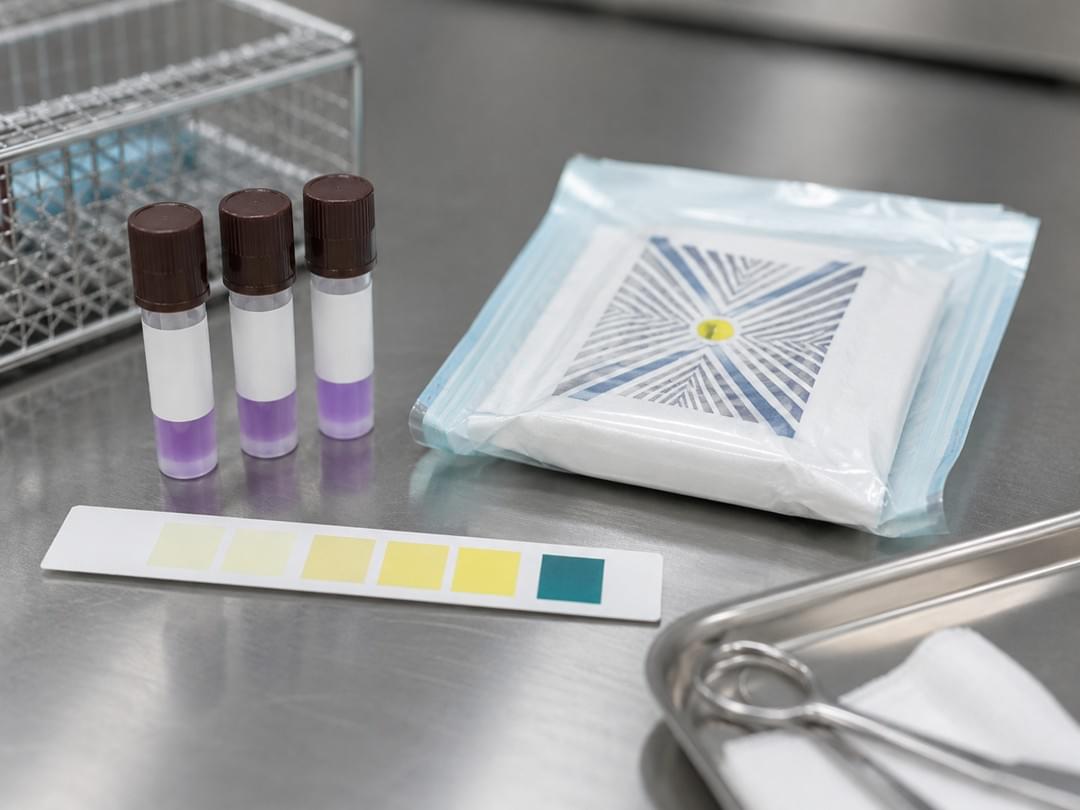
♨️ 5. Sterilization and Validation
Only after thorough cleaning, inspection, and packaging should instruments enter the sterilizer. Every sterilization cycle should be documented and verified using physical monitoring, chemical indicators, and scheduled biological testing to ensure consistent performance.
Skipping or rushing any of these stages weakens the entire infection control program. In practice, inadequate cleaning and poor packaging remain two of the most common causes of sterilization failure.
Veterinary Surgical Sterilization Stage 1: Ultrasonic Cleaning
Among all cleaning technologies, ultrasonic cleaners provide one of the most effective methods for removing contamination from complex surgical instruments.
Instead of relying on manual scrubbing alone, ultrasonic systems generate high-frequency sound waves—typically between 35 and 45 kHz—that create microscopic cavitation bubbles. As these bubbles collapse, they dislodge contaminants from surfaces that brushes often cannot reach.
Why Ultrasonic Cleaning Matters 🧪
Ultrasonic cleaning offers several important advantages:
- Removes debris from hinges, box locks, serrations, and narrow lumens
- Significantly lowers bioburden before sterilization
- Improves steam penetration during autoclaving
- Reduces manual labor while improving cleaning consistency
- Minimizes abrasive wear that can shorten instrument lifespan
Because sterilization can only be effective on thoroughly cleaned instruments, ultrasonic cleaning has become a standard component of modern veterinary reprocessing workflows.
Recommended Specifications
When selecting an ultrasonic cleaner, consider the following specifications:
| Feature | Recommendation |
| Tank Capacity | 3–6 L for general practices; 9 L or more for high-volume hospitals |
| Frequency | 35–45 kHz |
| Heating | 40–65°C heated cleaning solution |
| Lid Design | Fully enclosed to reduce aerosol exposure |
Although ultrasonic cleaning is highly effective, it should complement—not replace—proper rinsing, inspection, and routine maintenance.
Veterinary Surgical Sterilization Stage 2: Automated Washer-Disinfectors
As surgical caseloads increase, manual cleaning alone often becomes difficult to standardize. Automated washer-disinfectors help veterinary hospitals achieve more consistent, repeatable cleaning while reducing human error.
A complete cycle typically includes:
- ✅ Pre-rinse
- ✅ Enzymatic washing
- ✅ High-pressure rinsing
- ✅ Thermal disinfection
- ✅ Controlled drying
Because each cycle is automatically monitored, washer-disinfectors also generate digital or printed records that support audits and quality assurance.
Many validated systems comply with the ISO 15883 washer-disinfector standard, making them an excellent choice for hospitals seeking standardized instrument reprocessing.
For many practices, investing in automated washing becomes increasingly cost-effective once surgical volume reaches approximately 15 procedures per week. Beyond improving efficiency, automation also helps maintain consistent cleaning quality across different staff members.
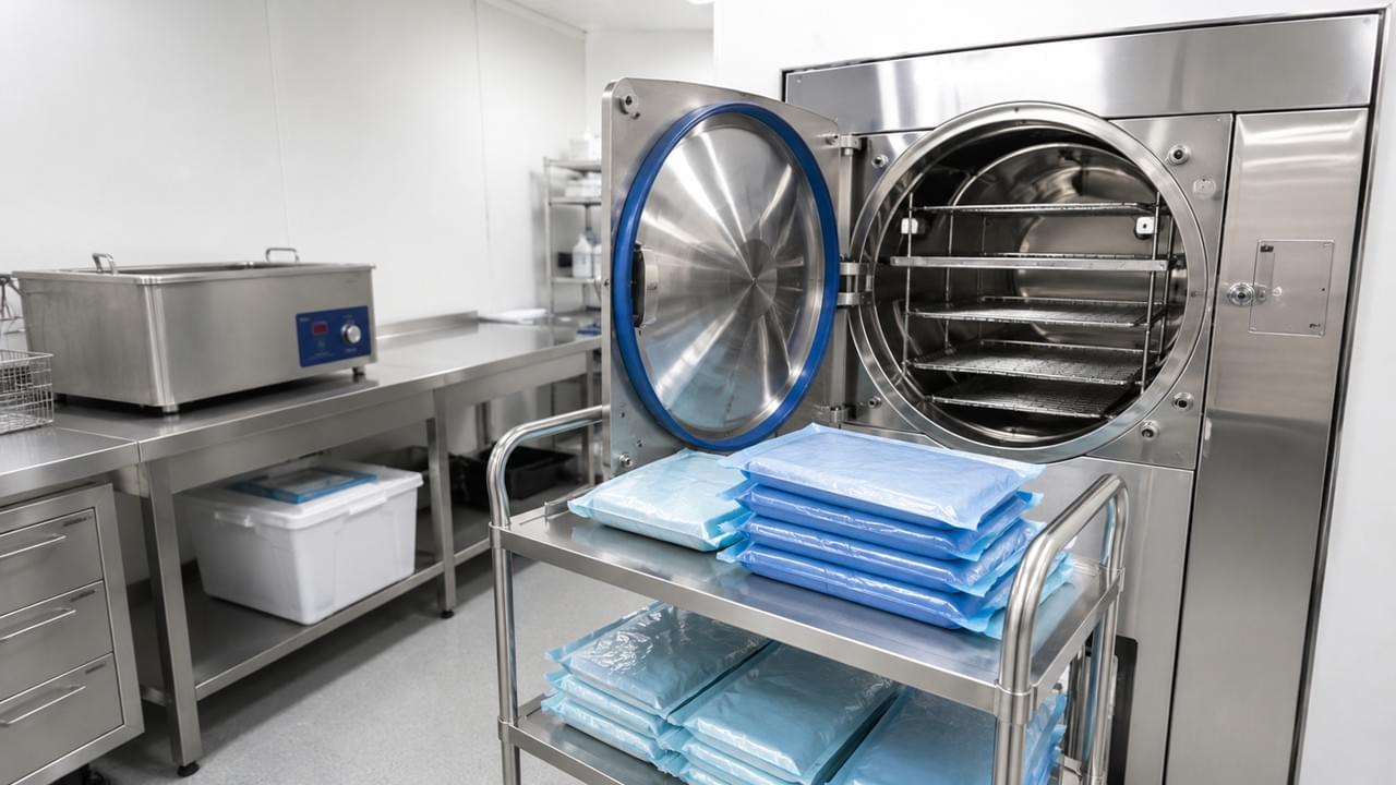
Veterinary Surgical Sterilization Stage 3: Choosing the Right Class B Autoclave
The autoclave remains the heart of every sterilization program. However, not all sterilizers deliver the same performance.
For modern veterinary hospitals, a Class B autoclave for veterinary clinics is widely recognized as the preferred solution because it is validated for challenging surgical loads, including:
- Wrapped surgical instrument trays
- Hollow instruments
- Cannulated devices
- Fabric packs
- Single- and double-wrapped surgical sets
Unlike Class N sterilizers, Class B systems use pre-vacuum technology that removes trapped air before steam enters the chamber. This allows steam to penetrate complex instruments much more effectively, producing consistently reliable sterilization results.
Why Class B Matters 🔬
Choosing an appropriate sterilizer directly affects patient safety. Class B systems provide reliable sterilization for the diverse instrument sets commonly used in orthopedic, soft tissue, dental, and minimally invasive veterinary procedures.
Clinics that routinely process wrapped surgical packs should consider Class B equipment the benchmark for best practice rather than an optional upgrade.
Validating Every Sterilization Cycle
Even the most advanced autoclave requires ongoing validation. Effective quality assurance should include:
| Validation Method | Recommended Frequency | Purpose |
| Physical cycle monitoring | Every cycle | Verify time, temperature, and pressure |
| Chemical indicators | Every package | Confirm steam penetration |
| Biological indicators | At least weekly | Verify sterilization using resistant bacterial spores |
| Bowie-Dick testing | Daily (Class B units) | Confirm efficient air removal and steam penetration |
Routine validation transforms sterilization from a simple mechanical process into a documented quality management system that supports patient safety, accreditation, and long-term operational excellence.
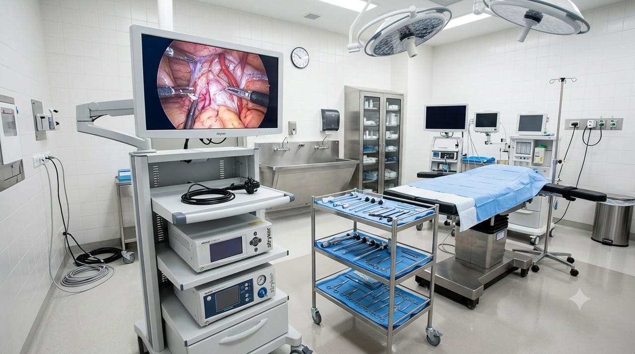
Veterinary Surgical Sterilization Stage 4: Infection Control Beyond Instrument Sterilization
Successful veterinary surgical sterilization extends far beyond instrument processing. 🏥 Even perfectly sterilized instruments can become contaminated if the operating room environment, staff practices, or patient preparation are not carefully controlled.
An effective infection prevention program combines environmental hygiene, controlled airflow, disciplined surgical techniques, and continuous staff training. When these elements work together, veterinary hospitals can significantly reduce the risk of surgical site infections while creating a safer environment for both patients and clinical teams.
If you’re planning or upgrading your surgical facility, our guide to How to Design an Efficient Veterinary Operating Room explores operating room layouts, workflow optimization, and equipment placement that further support infection prevention.
https://www.veteqpt.com/veterinary-operating-room-design-guide/
🧼 Surface Decontamination
Operating room surfaces become contaminated throughout the day through airborne particles, instrument handling, and staff movement. Consistent cleaning protocols help interrupt the transmission of microorganisms between procedures.
Between every surgery, staff should disinfect all frequently touched surfaces, including:
- Surgical tables
- Anesthesia machines
- Patient monitors
- Mayo stands
- Instrument carts
- Surgical light handles
- Door handles and switches
At the end of each working day, a comprehensive terminal cleaning should include floors, walls where appropriate, storage areas, and all high-touch equipment using veterinary-approved hospital disinfectants.
Rather than simply cleaning when contamination is visible, successful hospitals rely on standardized cleaning schedules that ensure every area receives consistent attention.
🌬️ Air Quality Management
Airborne contamination is often overlooked, yet it remains one of the most important contributors to postoperative infection.
Modern veterinary operating rooms should be designed to maintain clean airflow throughout every procedure. Recommended environmental practices include:
- HEPA-filtered ventilation systems
- Positive-pressure operating rooms
- At least 15 air changes per hour (ACH)
- Routine HVAC maintenance and filter replacement
- Continuous temperature and humidity monitoring where appropriate
Keeping operating room doors closed during procedures is equally important. Every unnecessary door opening disturbs airflow patterns and introduces unfiltered air that may carry bacteria and dust into the sterile field.
Limiting personnel traffic also reduces airborne particles. Studies consistently show that each additional person in the operating room increases microbial contamination within the environment.
🩺 Surgical Team Aseptic Technique
Even the best sterilization equipment cannot compensate for poor surgical discipline. Every member of the surgical team plays an essential role in maintaining sterility throughout the procedure.
Best practices include:
- Performing surgical hand preparation according to validated protocols
- Wearing properly fitted sterile gowns and gloves
- Using sterile draping techniques before instrument placement
- Avoiding unnecessary movement around the sterile field
- Replacing contaminated gloves immediately
- Maintaining a clear separation between sterile and non-sterile areas
Many veterinary hospitals now use alcohol-based surgical hand rubs as part of validated hand preparation protocols, provided they are applied correctly and only when hands are free of visible contamination.
Consistent training and periodic competency assessments help ensure these practices remain part of daily clinical routines rather than occasional reminders.
Documentation and Quality Compliance
📋 Effective veterinary surgical sterilization depends on more than completing each sterilization cycle—it also requires complete documentation. Accurate records provide traceability, demonstrate regulatory compliance, and help identify recurring issues before they affect patient outcomes.
Every veterinary practice should maintain documentation for:
- Sterilization cycle parameters
- Load identification numbers
- Chemical indicator results
- Biological monitoring records
- Instrument maintenance logs
- Equipment servicing reports
- Staff training records
- Surgical site infection surveillance
Comprehensive documentation also simplifies internal audits and supports accreditation programs by providing clear evidence that sterilization protocols are being followed consistently.
Continuous Quality Improvement
Sterilization programs should be reviewed regularly rather than treated as static procedures.
Routine quality reviews may include:
✅ Reviewing sterilization logs
✅ Monitoring biological indicator performance
✅ Inspecting instrument damage trends
✅ Auditing cleaning procedures
✅ Updating protocols after new equipment installation
✅ Staff refresher training
Small improvements implemented consistently often produce significant long-term reductions in infection risk while extending the service life of valuable surgical instruments.
Following International Best Practices
Veterinary hospitals should base their sterilization procedures on internationally recognized guidance whenever possible.
The WSAVA Infection Control Guidelines recommend maintaining documented sterilization records, monitoring postoperative infections, and establishing standardized cleaning procedures throughout the hospital. These recommendations help practices build reliable quality systems regardless of their size.
Similarly, practices using automated washer-disinfectors should select systems designed to meet the ISO 15883 washer-disinfector standard, ensuring validated and repeatable cleaning performance before sterilization.
For broader hospital hygiene strategies beyond the operating room, you may also find our guide to Infection Control & Disinfection Solutions in Veterinary Hospitals helpful:
https://www.veteqpt.com/infection-control-disinfection-veterinary-hospitals
Frequently Asked Questions
❓ Is a Class B autoclave necessary for every veterinary clinic?
Not every jurisdiction legally requires a Class B autoclave, but it is widely regarded as the best practice for practices that routinely sterilize wrapped instrument packs or hollow surgical instruments. Compared with Class N units, Class B sterilizers provide superior air removal and steam penetration, making them far more reliable for complex surgical loads.
❓ How often should surgical instruments be inspected?
Every instrument should be inspected during each reprocessing cycle before packaging. Staff should check for corrosion, damaged tips, loose joints, worn cutting edges, and alignment issues. In addition, high-value surgical instruments should undergo professional servicing at least once a year to maintain optimal performance.
❓ Can alcohol-based surgical hand rub replace traditional scrubbing?
Yes, validated alcohol-based surgical hand rubs can provide protection comparable to traditional surgical scrubbing when used correctly. However, hands must first be free of visible dirt or organic material, and manufacturers’ instructions regarding application volume and contact time should always be followed.
❓ What is the most common cause of sterilization failure?
In most veterinary hospitals, sterilization failures begin before the autoclave. Inadequate cleaning, improper packaging, overloaded sterilizers, and poor documentation are all common factors that compromise sterilization effectiveness. A standardized workflow with routine validation significantly reduces these risks.
🩺 Effective veterinary surgical sterilization is not defined by a single machine or procedure—it is the result of an integrated quality system that combines meticulous instrument reprocessing, validated sterilization equipment, environmental infection control, comprehensive documentation, and continuous staff training.
By investing in standardized workflows, choosing a Class B autoclave for veterinary clinics when appropriate, and following internationally recognized infection prevention practices, veterinary hospitals can improve surgical outcomes, protect patients, and build long-term confidence in the quality of their clinical care.
Whether you are establishing a new surgical suite or refining an existing sterilization program, every improvement made today contributes to safer surgeries tomorrow.