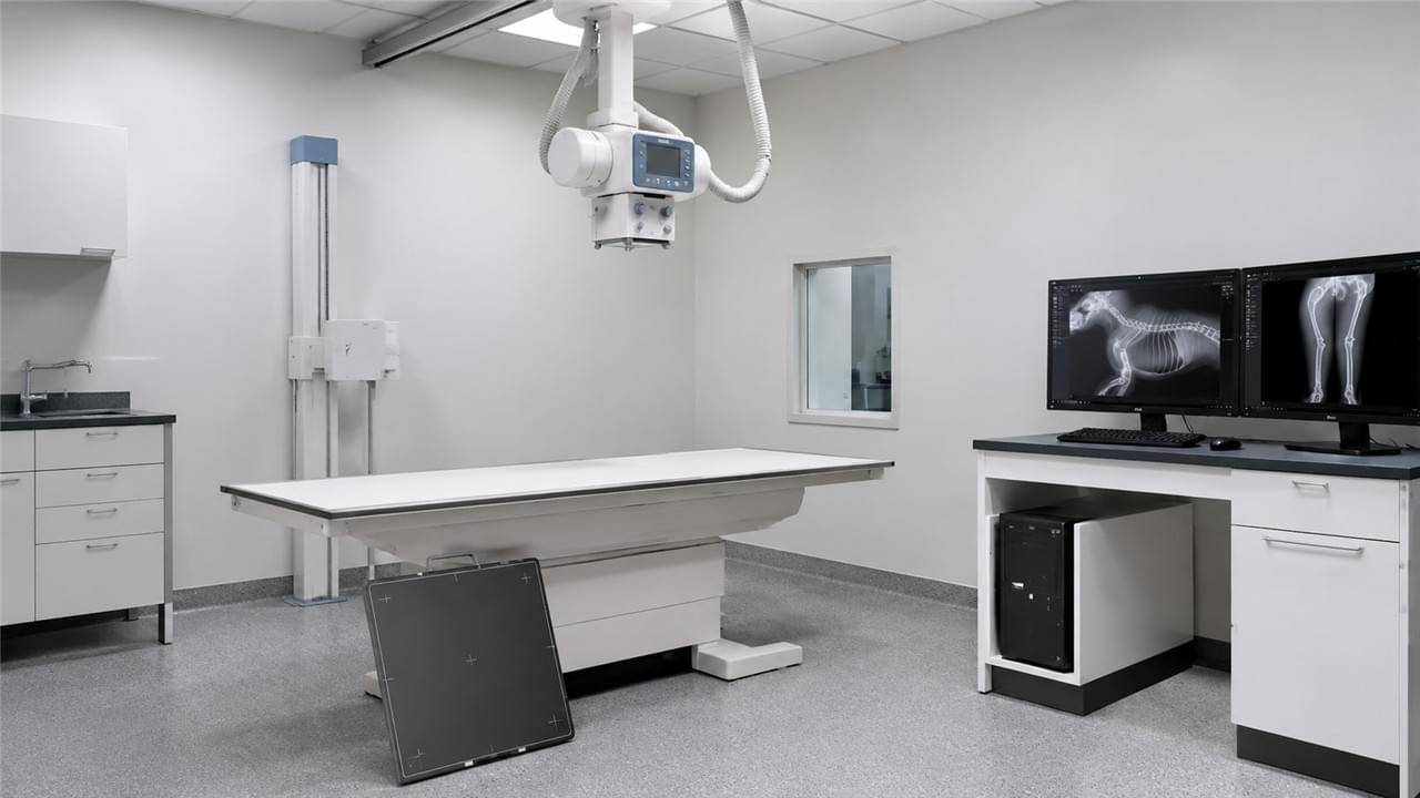
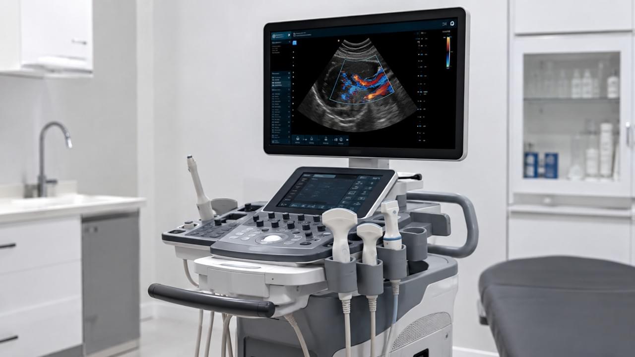
Veterinary digital X-ray systems have made film radiography obsolete — and computed radiography (CR) is next out the door. Flat-panel detectors capture and display images in 3–5 seconds, with better image quality at lower patient dose than anything that came before. It’s no longer a question of whether to go digital — it’s how to choose wisely.
Choosing digital X-ray systems for veterinary clinics means wading through detector technology, image quality metrics, software capability, installation requirements, and total cost of ownership — a stack of technical variables vendors love to drown you in. This guide cuts through the noise with clear, actionable guidance at every decision point. For the full diagnostic equipment context, see our guide on Veterinary Diagnostic Equipment: From Basic Exams to Advanced Imaging.
📡 DR vs. CR: Why Direct Digital Has Won
Why Veterinary Digital X-Ray Systems Beat CR Every Time
Computed radiography (CR) uses photostimulable phosphor plates that are scanned after exposure in a separate reader. It was a genuine upgrade over film when it arrived, but it has real limitations:
- Image readout takes 45–90 seconds per plate
- Plates degrade over time and need replacement
- Higher radiation dose required versus DR for equivalent image quality
Direct digital radiography (DR) with flat-panel detectors captures and displays the image in 3–5 seconds. No plate handling, no reader, no scan time. Modern DR systems deliver:
- Immediate image display on the clinical workstation
- Superior DQE (detective quantum efficiency) — better image quality at lower patient dose
- Lower long-term running cost (no replacement plates)
For any new installation in 2026, DR is the correct choice — and the gap between the two technologies only widens as veterinary digital X-ray systems mature.
🔬 Key Technical Specifications to Evaluate
The Specs That Define Veterinary Digital X-Ray Systems
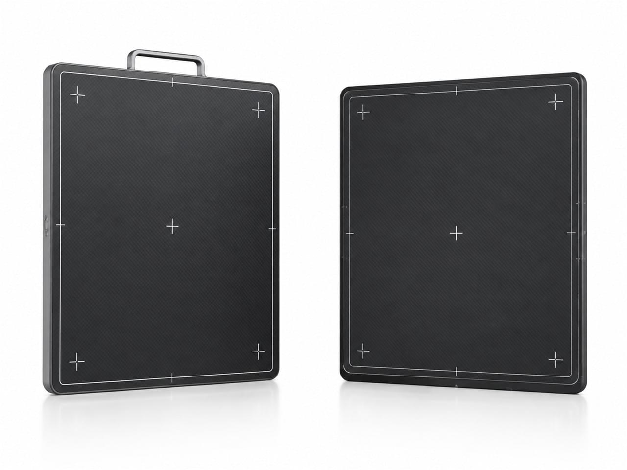
The detector is the heart of veterinary digital X-ray systems — scrutinize it hardest:
Detector Size
- 14×17″ (35×43 cm): standard for small/medium animal general practice
- 17×17″ (43×43 cm): useful for large breed dogs and equine practices
- 10×12″ (25×30 cm): supplementary small detector for extremity or dental work
Pixel Pitch and Spatial Resolution
- Pixel pitch: 100–150 microns is the current standard for veterinary DR
- Lower pixel pitch = higher inherent resolution (better bone detail, small patient imaging)
- Don’t over-specify resolution for soft tissue cases — match resolution to your actual clinical use
Detective Quantum Efficiency (DQE)
- DQE measures how efficiently the detector converts X-ray photons into image signal
- Higher DQE = better image quality at lower radiation dose
- Target: ≥60% DQE at 0 lp/mm for a quality system — this single number tells you more than any marketing slide
Detector Type: CsI vs. GOS
- CsI (cesium iodide): structured needle crystals absorb X-rays with minimal lateral light spread — higher DQE, better contrast resolution. The preferred choice when clinical excellence matters.
- GOS (gadolinium oxysulfide): unstructured phosphor — lower cost, slightly lower DQE. Acceptable when budget calls the shots.
Wireless vs. Tethered Detectors
- Wireless: maximum positioning flexibility — essential for large animal or field use, and a lifesaver for difficult small animal positioning
- Tethered (wired): lower cost, no battery management, no wireless interference risk — fine for a fixed table setup
Choosing the right veterinary DR flat-panel detector for your caseload comes down to these trade-offs — there’s no universal answer, only the right answer for your practice.
💻 Software and DICOM Compliance
Software: The Half of Veterinary Digital X-Ray Systems Everyone Forgets
Image acquisition hardware is only half the system. The software platform determines usability, efficiency, and integration — and it’s where most practices feel buyer’s remorse.
Essential software features:
- Veterinary DICOM acquisition and storage
- Patient registration with species, breed, and body part selection
- Automatic image processing with species-specific algorithms (bone vs. soft tissue window presets)
- PACS integration (DICOM send to server or cloud)
- Multi-image comparison view (current vs. prior study)
- Measurement and annotation tools
Integration checklist — ask these three questions before you sign:
- Does the system export standard DICOM 3.0?
- Can it integrate with your practice management software (ezyVet, Idexx Neo, VetSmart)?
- Does it support DICOM worklist (auto-populating patient data from the PMS to eliminate manual entry errors)?
Software is where veterinary digital X-ray systems either earn their keep or frustrate you daily. Don’t let a demo on a sales laptop fool you — ask for a test drive on your own workstation.
☢️ Radiation Safety Requirements for DR Installation
Radiation Safety: The Non-Negotiable Side of Veterinary Digital X-Ray Systems
Digital X-ray systems need a properly constructed room regardless of digital format — the radiation source is the X-ray tube, unchanged from the film era.
Key installation requirements:
- Lead shielding on walls, floor, and ceiling (verified by a radiation physicist’s calculation based on expected workload)
- Operator shielding (lead glass window or remote exposure from the corridor)
- Radiation safety signage (“X-ray in use” indicator)
- Annual QA testing of X-ray tube output and exposure accuracy
- Personal dosimetry for operators per local regulatory requirements
Consult a qualified medical radiation physicist before room construction — the AVMA radiation safety resources provide a useful reference framework. Cutting corners on shielding isn’t a budget win; it’s a liability you’ll carry for the life of the room.
💰 Total Cost of Ownership: Beyond the Sticker Price
The Real Cost of Veterinary Digital X-Ray Systems
| Cost Component | Typical Range |
|---|---|
| System purchase (DR system + software) | 18,000–18,000–55,000 |
| Room construction (shielding) | 5,000–5,000–20,000 |
| Installation and commissioning | 2,000–2,000–5,000 |
| Annual software license | 500–500–2,000/year |
| Annual maintenance contract | 1,500–1,500–4,000/year |
| Detector replacement (every 7–10 years) | 8,000–8,000–18,000 |
Total 5-year TCO for a standard veterinary DR installation: roughly 40,000–40,000–90,000 depending on specification and market. That’s why the sticker price is the least interesting number on this page — the recurring costs decide the real economics of veterinary digital X-ray systems.
❓ Frequently Asked Questions
Q: Can I retrofit a digital X-ray detector onto my existing analogue X-ray generator?
A: In many cases, yes — and it’s the cheapest route into veterinary digital X-ray systems if your generator meets minimum output specifications (typically 40–150 kVp, 10–500 mAs range). Retrofit detectors are designed to work with existing generators, significantly reducing cost by reusing what you already own. But a radiation safety assessment is required first: the generator’s exposure accuracy and reproducibility must be verified before a DR retrofit for veterinary clinics makes clinical sense.
Q: What is the typical lifespan of a veterinary DR flat-panel detector?
A: Most flat-panel detectors are rated for 200,000–400,000 exposures, which translates to 7–12 years at moderate clinic volume. Wireless detectors with battery systems may need battery replacement at 3–5 years. Annual QA testing tracks performance degradation and signals when replacement is approaching — a routine reality of owning veterinary digital X-ray systems.
Q: Is dental X-ray separate from general radiography, or can the same DR system serve both?
A: They’re separate systems. General radiography DR systems use large-format detectors optimized for thorax, abdomen, and limb imaging. Dental radiography requires small-format intraoral sensors or phosphor plates positioned inside the patient’s mouth for periapical imaging. These are inherently different hardware configurations — budget for them separately.
✅ The Bottom Line
Buying veterinary digital X-ray systems doesn’t need to feel like a physics exam. Prioritize detector quality (DQE), demand software that integrates with your workflow, budget for the room and the recurring costs, and verify radiation safety before anything else. The WSAVA diagnostic imaging standards are a good place to ground your expectations. And if you’re building out the full imaging suite, see how X-ray fits alongside our guide on How to Choose Veterinary Ultrasound Equipment.