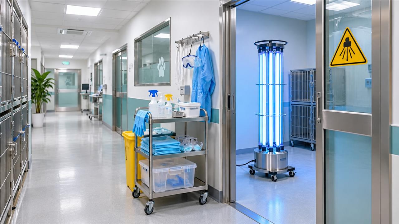
Nosocomial infections — healthcare-acquired infections — are a recognized and significant risk in veterinary hospital environments. Methicillin-resistant Staphylococcus pseudintermedius (MRSP), multidrug-resistant gram-negative organisms, and highly contagious viral diseases such as parvovirus and panleukopenia can persist in inadequately disinfected environments and cause preventable patient harm.
Effective veterinary infection control is a systems-level discipline: physical isolation infrastructure, correct disinfectant selection and application, validated sterilization, air management, and staff hygiene protocols — working together. No single product or gadget does the job. This guide covers all seven dimensions of infection control and disinfection solutions in veterinary hospitals.
For the foundational setup context, refer to our Complete Guide to Veterinary Hospital Equipment Setup.
🔗 The Infection Control Chain: Understanding the Risk Points
🚦 The Four Transmission Routes Veterinary Infection Control Must Block
Infectious agent transmission in a hospital occurs through four pathways — map them first, because every veterinary infection control decision downstream answers one of them:
- 🤝 Direct contact: patient-to-patient contact, particularly in group housing
- 🖐️ Indirect contact (fomites): contaminated surfaces, equipment, hands, clothing
- 💨 Aerosol/droplet: respiratory pathogens — Bordetella, influenza, SARS-CoV-2 in feline populations
- 🐜 Vector: ectoparasites and fomite-borne transmission on staff clothing
Sound veterinary infection control addresses all four pathways simultaneously. No single disinfectant or piece of equipment prevents every route — which is why the seven systems in this guide overlap deliberately.
Fomite transmission deserves special respect because it is the route staff enable without noticing. A stethoscope moved from a parvovirus puppy to a post-operative spay, a jacket sleeve brushed along a cage bank, a phone checked between patients — each is a plausible vector. Environmental sampling studies in veterinary clinics routinely recover pathogenic organisms from door handles, computer keyboards, and the floor areas around kennels, which is exactly why the systems below pair physical infrastructure with procedural discipline.
The economics also justify the engineering: a single MRSP cluster can shut down an operating schedule for days, a parvovirus contamination event can force ward closure and environmental decontamination costing thousands, and one documented nosocomial outbreak can undo years of client trust. Infection control spending looks cheap measured against any of those outcomes. 💸
🏥 Infrastructure: Isolation Room Design and Equipment
🚪 Isolation Rooms: The Cornerstone of Veterinary Infection Control
The physical isolation room is the most critical veterinary infection control infrastructure investment for practices managing contagious patients. See our dedicated guide: Veterinary Isolation Rooms: Design and Equipment Guide.
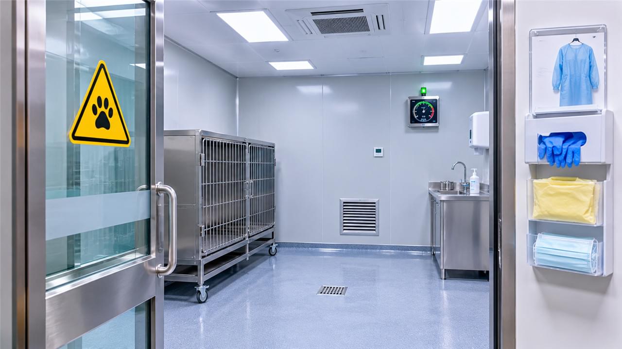
Minimum veterinary isolation room design requirements:
- 🔒 Physically separate room with self-closing door — not a curtained-off area
- 🌬️ Negative pressure ventilation (air exhausted outside, never recirculated) OR independently exhausted HEPA-filtered recirculation
- 🧴 Dedicated disinfection supplies that never leave the isolation room
- 🥼 Dedicated PPE storage at the room entrance
- 🚰 Clinical sink with elbow-operated or touchless faucet inside the room
- 🕳️ Floor drain for liquid waste management
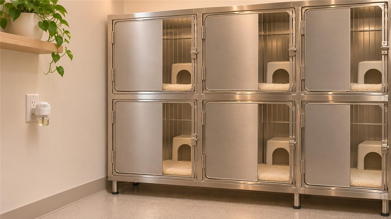
Isolation cage requirements: a solid wall panel separating from adjacent non-isolation cages, individual drainage independent of the main ward drain system, and lining in an easily disinfectable material.
Operating the room well matters as much as building it: assign dedicated staff who enter only when needed, don and doff PPE in a fixed sequence at the entrance, keep a log of every entry and exit, and route consumables through a single supply point or pass-through. Foot traffic is an aerosol pump — every unnecessary visit re-pressurizes the doorway and re-contaminates the corridor. Clinics that treat isolation as a staffing protocol rather than just a room are the ones that actually contain outbreaks.
For short-stay clinics that cannot justify a full isolation room, the intermediate standard is a dedicated isolation cage bank with solid barriers, separate drainage, and its own labeled cleaning kit — acceptable for brief holds of stable patients, never for aerosol-generating respiratory cases or active parvovirus. It buys time, but it is not a substitute for full veterinary infection control infrastructure.
🧪 Disinfectant Selection: Choosing the Right Product for Each Situation
🧯 Building the Disinfectant Formulary for Veterinary Infection Control
No single disinfectant is effective against all pathogens at all concentrations — and disinfectant selection is the daily core of veterinary infection control. A hospital should maintain a formulary of 2–3 disinfectants with complementary spectra:
| Disinfectant Class | Spectrum | Common Veterinary Use |
|---|---|---|
| Accelerated hydrogen peroxide (AHP) | Broad-spectrum including parvovirus, spores | General surface disinfection, isolation rooms |
| Quaternary ammonium compounds (QUAT) | Bacteria, enveloped viruses; NOT parvovirus | General ward disinfection |
| Sodium hypochlorite (bleach) | Broad including non-enveloped viruses, spores | Parvovirus/panleukopenia decontamination |
| Potassium peroxymonosulfate (Virkon-S type) | Broad-spectrum including non-enveloped viruses | Isolation room terminal disinfection |
| Phenolics | Bacteria, mycobacteria, fungi | NOT safe for cats — avoid in feline areas |
⏱️ Contact time is critical: no disinfectant works below its minimum contact time. Apply at the correct dilution, keep the surface visibly wet for the specified period (typically 2–10 minutes), then let it dry. Spray-and-immediately-wipe is disinfection theater — the label contact time is the product.
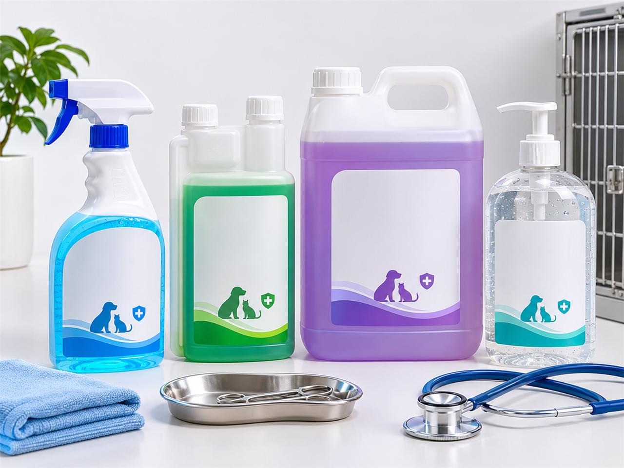
Practical formulary notes for veterinary infection control: verify dilution with dosing pumps or pre-measured concentrates — eyeballed bleach dilutions routinely land outside the effective range. Check material compatibility: concentrated bleach corrodes stainless finishes and degrades rubber seals. Label every spray bottle with contents, dilution date, and required contact time so a relief nurse can run the system correctly at 11 p.m. Rotate actives only when resistance or compatibility concerns justify it — consistency beats novelty in disinfection chemistry.
⚡ Equipment-Based Disinfection: UV vs. Ozone Technologies
🔦 Automated Disinfection Technologies in Veterinary Infection Control
Beyond manual chemical methods, automated technologies extend veterinary infection control to whole room volumes with consistent, repeatable results. For a detailed comparison, see UV vs Ozone Disinfection in Veterinary Clinics.
UV-C disinfection units:
- 💡 UV-C (254 nm) disrupts pathogen DNA/RNA — inactivating bacteria, viruses, and fungi
- 🛋️ Effective for surface and air disinfection in unoccupied rooms
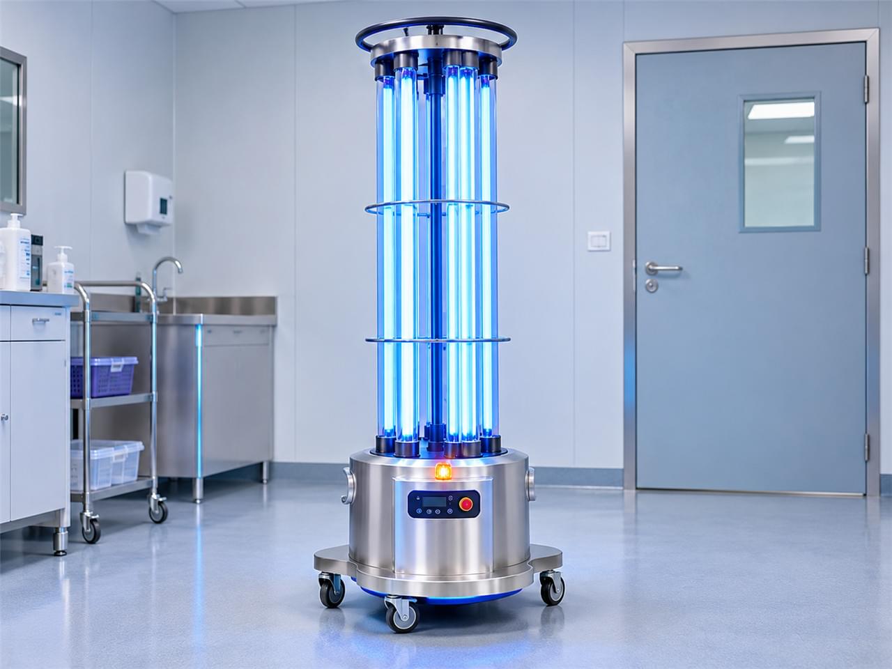
- 🗼 Mobile tower units handle terminal room disinfection after infectious patients
- ✅ No chemical residue — safe for patient re-entry after a standard ventilation period
- ⚠️ Limitation: shadows create protected zones — cage corners and furniture out of line-of-sight receive a reduced dose
Ozone generators:
- 🌪️ High-concentration ozone (O₃) is a powerful oxidant inactivating a broad pathogen range
- 🧭 More penetrating than UV — reaches surfaces behind furniture and into corners
- ⛔ Safety first: the room must be evacuated (humans and animals) during treatment, and ozone must fully dissipate before re-entry — minimum 2 hours, verified with an ozone detector
Running a UV-C terminal cycle well: clear the room completely, position the tower centrally with line-of-sight to as many surfaces as possible, open cage doors and drawers so shadowed interiors receive direct exposure, set the timer per the manufacturer’s dose guidance for the room volume, and post a warning sign at the door while it runs. This is veterinary infection control at its most mechanical — the machine works brilliantly on what it can see, and your job is to hide nothing from it. UV-C’s shadow limitation and ozone’s penetration advantage are two halves of the same trade-off, and larger facilities often run both.
Ozone’s penetration, meanwhile, trades directly against its re-entry burden: budget the full two-hour dissipation plus detector verification into the discharge-cleaning workflow, or schedule pressure will tempt the shortcuts that defeat the technology’s purpose.
🔧 Instrument Reprocessing: Washer-Disinfectors
🫧 Washer-Disinfectors: Validated Cleaning Inside Veterinary Infection Control
Washer-disinfectors provide automated, validated instrument cleaning — a pillar of veterinary infection control that manual washing cannot match. For the full walkthrough, see Washer-Disinfectors in Veterinary Instrument Reprocessing.
Key validation elements:
- 📋 Cycle parameter documentation — temperature, time, detergent concentration
- 🧫 Periodic protein residue tests verifying actual cleaning efficacy
- 🔧 Annual service and calibration
The word that matters is validated: a manual sink tells you nothing about what actually happened inside a locked hemostat box.
A practical purchasing note: match the chamber size to your heaviest instrument volume, not your average day — the unit that runs three cycles every evening is the unit that quietly becomes the bottleneck that reintroduces hand-washing habits. Thermal disinfection phases (typically 90–93°C) add a verified kill step that a sink can never offer, and cycle printouts give your sterilization log the documentation auditors and insurance assessors actually want to see.
🛡️ Preventing Cross-Contamination: Procedural and Equipment Strategies
🚫 Cross-Contamination in Veterinary Hospitals: The Daily Defenses
Cross-contamination in veterinary hospitals happens in the boring moments — the shared stethoscope, the mop bucket that tours every ward. Dedicated guidance: Preventing Cross-Contamination in Veterinary Hospitals. These defenses are where veterinary infection control stops being infrastructure and becomes daily habit:
Key strategies:
- 🏷️ Dedicated equipment per patient: IV poles, monitoring equipment, and fluid bags labeled per patient — never shared during active use
- 🎨 Color-coded cleaning systems: mop heads, cloths, and buckets per zone, keeping dirty (isolation) and clean (surgical) areas strictly separate
- 🧤 Single-use items where practical: surgical gloves, single-use blood collection sets, individual patient medication kits
- 🖐️ Hand hygiene at every transition: alcohol-based hand rub at every cage, every door, every shared equipment item
Put the schedule in writing: daily cleaning of all occupied cage fronts and high-touch surfaces, weekly deep-cleaning of each ward in rotation, and immediate spot-cleaning after any visible contamination. Zone your staff — nurses assigned to isolation do not float into surgery prep mid-shift without a full change of outerwear. Laundry and clinical waste leave the building on labeled, closed routes that never pass through clean areas. None of this is glamorous; all of it is what keeps cross-contamination in veterinary hospitals from becoming an outbreak investigation.
Sterile processing upstream belongs to Sterilization & Infection Control in Veterinary Surgical Suites — this ward-level layer catches everything that moves between patients.
📋 Biosecurity Standards in Modern Veterinary Facilities
🌐 Biosecurity: The Framework Behind Veterinary Infection Control
For the comprehensive framework, see Biosecurity Standards in Modern Veterinary Facilities. The WSAVA infection control guidelines provide the internationally referenced standard for companion animal facilities, with AVMA biosecurity and infection control standards complementing them at the policy level. Together they turn veterinary infection control from a set of purchases into a managed program. Key components:
- 🔍 Infectious disease triage protocol — suspect infectious patients handled at intake, not discovered in the ward
- 💉 Vaccination admission requirements — confirm status for all elective admissions
- 📊 Surveillance and recording — track healthcare-acquired infections, investigate clusters
- 🎓 Staff education and hand hygiene auditing
Biosecurity is veterinary infection control made routine: written, scheduled, audited — rather than heroic.
Each component earns its place. Triage at intake stops the parvo puppy from touring your waiting room. Vaccination checks protect the elective admissions already in the ward. Surveillance turns “it seems like we’ve had a few GI cases lately” into an investigated cluster with a source. And hand-hygiene auditing converts good intentions into measured behavior. Review the written protocols annually — organisms change, staff change, and a biosecurity plan that has not been revisited is a plan quietly going stale.
🧼 Staff hygiene deserves its own line in the budget: handwashing technique training at onboarding, glove discipline that distinguishes contact precautions from routine care, facility-provided uniforms laundered on site rather than worn home, and a culture where anyone — from kennel assistant to practice owner — can call out a missed hand hygiene step without awkwardness. The cheapest layer of the veterinary infection control stack is also the layer most dependent on leadership.
❓ Frequently Asked Questions
Q: What disinfectant kills parvovirus on hospital surfaces?
A: Canine parvovirus (CPV) and feline panleukopenia (FPV) are non-enveloped viruses with extreme environmental stability — surviving on surfaces for months. Effective options: sodium hypochlorite (bleach) at 1:30 dilution, accelerated hydrogen peroxide products (e.g., Accel, Rescue), and potassium peroxymonosulfate (Virkon-S). QUATs are NOT effective against parvovirus — the single most important disinfectant selection point in veterinary practice.
Q: How often should veterinary isolation rooms be terminally cleaned?
A: Terminal cleaning — complete chemical disinfection of all surfaces plus automated air disinfection where available — should follow every infectious patient discharge. The schedule is patient-driven, not time-driven, and that responsiveness is what keeps veterinary infection control ahead of an environmental reservoir. Additionally, terminal-clean the room weekly regardless of occupancy to prevent accumulation of resistant organisms.
Q: Is a dedicated isolation room necessary, or is a designated cage in the main ward adequate?
A: For any meaningful volume of respiratory, gastrointestinal, or highly contagious patients, a dedicated physical isolation room is strongly recommended — and required by practice regulations in most jurisdictions. A designated ward cage cannot provide adequate aerosol containment, hands-free traffic management, or independent drain/waste management — all necessary for true infectious disease isolation. A dedicated room remains the foundation of veterinary infection control for any serious contagious caseload.
📚 Related Articles in This Series
- Veterinary Isolation Rooms: Design and Equipment Guide
- UV vs Ozone Disinfection in Veterinary Clinics
- Washer-Disinfectors in Veterinary Instrument Reprocessing
- Preventing Cross-Contamination in Veterinary Hospitals
- Biosecurity Standards in Modern Veterinary Facilities
🎯 Key Takeaways
Veterinary infection control is seven overlapping systems, not one purchase: blocked transmission routes, a real isolation room, a complementary disinfectant formulary, automated UV or ozone terminal disinfection, validated instrument reprocessing, daily cross-contamination discipline, and a written biosecurity framework. Build the system before the outbreak forces you to — and keep the WSAVA infection control guidelines on the wall, not in a drawer. Your patients arrive healthy. The building’s job is to send them home that way. 🐾